A micro-focused x-ray source illuminates the object of interest, which is positioned on a precision manipulator. The x-ray shadow images are acquired by a sensitive x-ray camera. During the image acquisition the sample is rotated a step at a time through 180 degrees. Images are recorded at each rotation. Using complex software 2 dimentional images (or slices) based on x-ray density can be recalculated from the x-ray shadow images.
One or more of these 2D images can be stored in the computer and then using another software packages a stack of these images can be added together to form a 3D model of the sample. From this model it is possible to make virtual cuts or slices of the object in any direction. The software allows for numerical data to be calculated from the morphology of the samples structure.
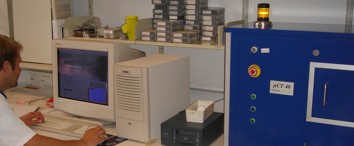
The lab has a microCT scanner, µCT 40 by Swiss Scanco. The machine can scan down to 9µm for a 10mm object, even at its full capacity the scanner uses a resolution around 20µm.The maximum size of an object for the microCT scanner is 36mm in diameter and 80 mm in length.
The lab also owns a µCT 20. For a detailed product description please visit Scanco, or stop by the lab to discuss the equipment in detail. |